Case · 01
Making functional brain maps clinically inspectable
Turning a black-box neuroimaging output into a workflow radiologists can inspect, adjust, and defend.
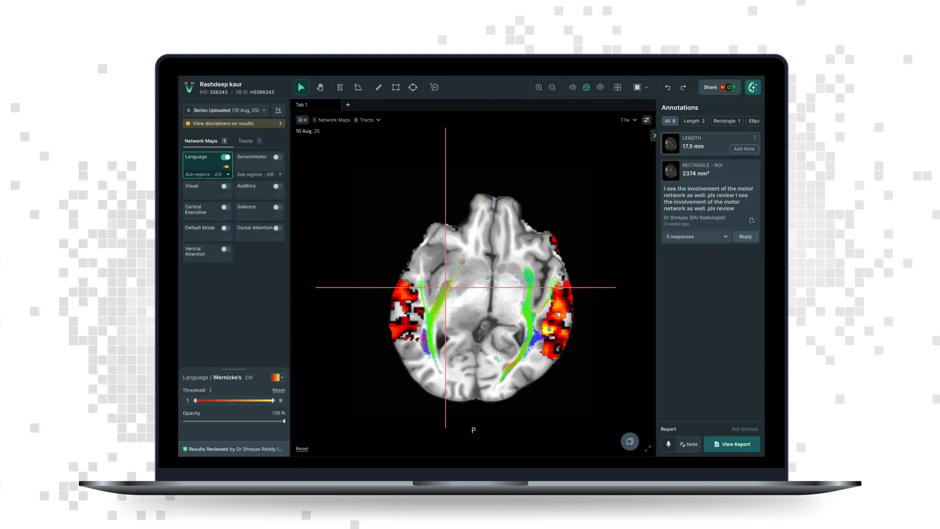
Neuroscience SaaSI led the design of VoxelBox, a clinical neuroimaging workspace that brings structural MRI, functional networks and white-matter tractography into one place.
The problem was not simply displaying more imaging data. Radiologists needed to decide whether the generated maps were anatomically credible, understand what could influence the surgical plan, and communicate that clearly to the neurosurgeon.
The problem
Functional maps can add important context to a structural MRI, but they are difficult to produce, unfamiliar to many radiologists and easy to treat as a black box.
The existing workflow placed the outputs next to the clinical process rather than inside it. Radiologists could view results, but had limited support for verifying, adjusting, comparing or carrying their interpretation into the report.
For the product to be useful, the output had to become inspectable.
My role
I led the product design across the viewer, working with radiologists, neuroscientists, engineering, product and regulatory teams.
My responsibility was to turn a technically complex set of outputs into a workflow that clinicians could understand and control without oversimplifying the underlying science.
The design model
I organised the experience around three actions.
Confirm
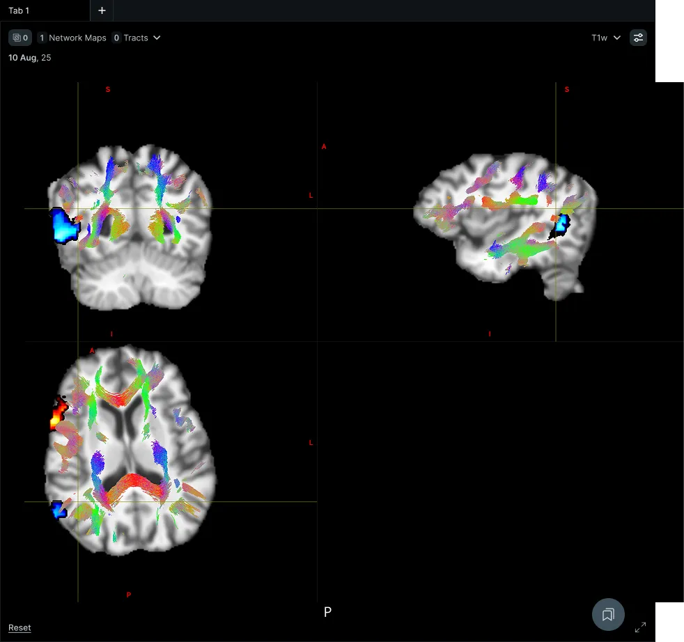
Help the radiologist determine whether the output is credible. This meant placing functional maps directly alongside structural anatomy, linking views through the crosshair, exposing relevant metrics and making it easier to inspect a result at both the whole-brain and local level.
Modify
Give the clinician meaningful control where interpretation required it. Radiologists could adjust thresholds, opacity and tract refinement while retaining a clear relationship with the generated result. The goal was not to let users freely redraw the science, but to help them examine it under different conditions.
Conclude
Turn the interpretation into something another clinician could use. Annotations, bookmarks, measurements and reporting were designed as part of the review workflow, rather than separate utilities added around the viewer.
Key decisions
Keep the anatomy primary
Functional networks and tracts were designed as overlays, not as standalone visualisations. The structural MRI remained the reference point throughout the workflow. This gave radiologists a familiar anchor and made the generated output easier to assess against anatomy.
Treat display controls as clinical controls
Opacity, thresholding and tract refinement can look like simple visual settings. In practice, they change what the clinician can see and therefore what they may conclude. I designed these controls to be easy to access while making their effect legible and reversible.
Connect global and local interpretation
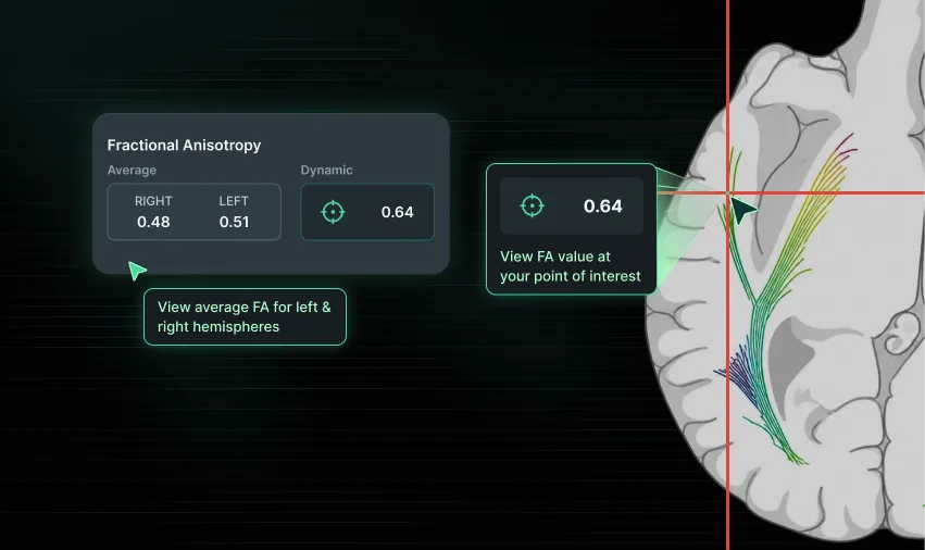
A tract-level value is useful, but it does not always explain what is happening at a specific anatomical location. We introduced both static left-versus-right tract values and values that update based on the crosshair position. This supported comparison at a summary level and closer inspection at the point of interest.
Preserve evidence during review
Measurements, annotations and bookmarks were designed to help radiologists retain findings while moving through multiple planes and overlays. They became a bridge between image interpretation and reporting, rather than a separate drawing toolset.
Design reporting as the end of review
The workflow allowed the radiologist to move from examining the scan to recording findings without leaving the clinical context. Notes, selected views and relevant findings could be carried into the report, reducing the need to reconstruct the interpretation afterwards.
What changed
VoxelBox moved from being a place where generated outputs could be viewed to a workspace where they could be examined, adjusted and reported.
The resulting product gave radiologists a more active role in functional brain mapping and created a clearer handoff to the neurosurgeon.
Why this work matters
AI output becomes useful in healthcare only when the responsible clinician can understand what they are looking at, inspect its limitations and communicate the conclusion.
The viewer was designed around that responsibility.